March 2020

Example of a neutron microtomography using the PSI 'Neutron Microscope’ at the ILL-D50 beamline: A vertical slice from a neutron microtomography dataset showing dendritic
microstructures of lead, voids and gold in a sample of a gold-lead alloy.
Figure published in the WCNR-11 paper:
"PSI ‘Neutron Microscope’ at ILL-D50 Beamline - First Results"
Pavel Trtik, Michael Meyer, Timon Wehmann, Alessandro Tengattini, Duncan Atkins, Eberhard Lehmann, Markus Strobl
Materials Research Proceedings 15 (2020) 23-28
Reproduced by permission of the authors.

April 2020
May 2020
Left: radiographic image of maltodextrin particles (x = 3.55 mm, c = 0.05 w/w) with the respective transmission scale;
Right: tomographic image of the maltodextrin particle (x = 3.55 mm, c = 0.05 w/w) at a height of 1.55 mm (red line in the left image) with the absorption scale (a.u.); the particle edge is represented by the red dotted line; the green line indicates the actual position of the drying front.
Figure published in the paper:-
Hilmer M, Peters J, Schulz M, Gruber S, Vorhauer N, Tsotsas E, Foerst P
Reproduced from The Review of Scientific Instruments, 01 Jan 2020, 91(1):014102, with the permission of AIP Publishing.
DOI: 10.1063/1.5126927 PMID: 32012547
Egyptian objects from the Kha and Merit grave goods.
Left: sealed ceramic vase investigated through neutron techniques, neutron radiography, and PGAA plot with the labels of the detected isotopes;
Right: Egyptian metallic vase (situla), neutron radiography and one of the acquired diffraction patterns
Figure published in the paper:
"Neutrons for Cultural Heritage—Techniques, Sensors, and Detection"
Giulia Festa , Giovanni Romanelli , Roberto Senesi, Laura Arcidiacono, Claudia Scatigno, Stewart F. Parker, M. P. M. Marques, Carla Andreani
Sensors 2020, 20(2), 502; https://doi.org/10.3390/s20020502
Reproduced with permission from G. Romanelli, Sensors; published by MDPI, 2020.

June 2020

Photo (a) and a fast neutron image (b) of the Li-fueled reactor, post-operation. The flat and dark-field corrected radiographic image reveals a heat exchanger coil, the upper highly oxidized region (dark-gray), and the lower, primarily non-oxidized, elemental Li-rich portion (light gray). The image was taken over four minutes of exposure time. The red line represent roughly the beam size at the detector position.
Figure published in the WCNR-11 paper:
"Fast Neutron Imaging at a Reactor Beam Line"
R. Zboray, Ch. Greer, A. Rattner, R. Adams, Z. Kis
Materials Research Proceedings 15 (2020) 180-184
Reproduced by permission of the authors.
July 2020

Organisation of electrode unrolling (top, middle, bottom) and analysis of the lithium distribution in the LixMnO2 electrode during discharging using neutron tomography data
Figure published in the paper:-
Ralf F. Ziesche, Tobias Arlt, Donal P. Finegan, Thomas M. M. Heenan,
Alessandro Tengattini, Daniel Baum, Nikolay Kardjilov, Henning Markötter,
Ingo Manke, Winfried Kockelmann, Dan J. L. Brett & Paul R. Shearing
Nature Communications volume 11, Article number: 777 (2020)
Reproduced with permission from P.R. Shearing
August 2020

Figure published in the paper:-
"Neutron Dark-Field Imaging with Edge Illumination"
Marco Endrizzi, Gibril K. Kallon, Triestino Minniti, Rolf Brönnimann, Alessandro Olivo
arXiv:2006.12171v1 [physics.ins-det] 22 Jun 2020
Reproduced with permission from Marco Endrizzi

September 2020
Figure 5
a) Neutron CT slice shows layers of textile wrapping;
b) X-ray CT slice shows higher grayscale contrast;
c) Segmented and visualised calcaneus bone [green area in b)];
d) Close-up showing layered wrapping of varying tightness and coarseness [red box on a)]
Figure published in the WCNR-11 paper:-
"Digitally Excavating the Hidden Secrets of an Egyptian
Animal Mummy: a Comparative Neutron and X-ray CT Study"
Carla A Raymond and Joseph J Bevitt
Materials Research Proceedings 15 (2020) 250-255
Reproduced by permission of the authors.
October 2020


Figure published in:-
"Electric field imaging using polarized neutrons"
Yuan-Yu Jau, Daniel S. Hussey, Thomas R. Gentile, Wangchun Chen
arXiv:2006.03728 [physics.ins-det]
Reproduced by permission of the authors

November 2020
Figure 7. X-μCT- (A) and n-μCT-based (B) mesiodistal virtual sections through the buccal cusps of the lower molar SMF-8888. The white arrows indicate the positions of two pulp horns barely discernible on the X-ray image (A) but clearly rendered by the neutron-based record (B). The dotted line in (B) highlights the enamel-dentine boundary, not visible in (A).
Figure published in:-
"When X-Rays Do Not Work. Characterizing the Internal Structure of Fossil Hominid Dentognathic Remains Using High-Resolution Neutron Microtomographic Imaging"
Clément Zanolli, Burkhard Schillinger, Ottmar Kullmer, Friedemann Schrenk, Jay Kelley, Gertrud E. Rössner and Roberto Macchiarelli
Front. Ecol. Evol., 27 February 2020 https://doi.org/10.3389/fevo.2020.00042
Reproduced by permission of Clément Zanolli
December 2020


Figure 4.
(a) The horizontal cut through the tomography image of the partially dried sample (after 11.5 h of drying) reveals the dry zones. The light-gray regions contain water and maltodextrin (it is noted that maltodextrin and heavy water are not distinguished from this image).
(b) Schematic illustration of the different zones that are distinguished from the tomography image. The red arrows depict the expansion direction of the dry zones.
1: accumulation of maltodextrin inside the peripheral maltodextrin belt;
2: peripheral sublimation zone with fractal front;
3: ring of frozen water and maltodextrin;
4: sublimation fingering zone.
Figure published in:-
"Freeze-Drying with Structured Sublimation Fronts—Visualization with Neutron Imaging"
Nicole Vorhauer-Huget, David Mannes, Mathias Hilmer, Sebastian Gruber, Markus Strobl, Evangelos Tsotsas, Petra Foerst
Processes 2020, 8(9), 1091
https://doi.org/10.3390/pr8091091
Reproduced by permission of Nicole Vorhauer-Huget

January 2021
Fig. 8. NCT-rendered images of pristine (room temperature) and pyrolyzed (1000 °C) samples: (a) axial and (b) radial throughcuts of the samples defined in Table 2 and shown in Fig. 9.
Figure published in "Dynamics of hydrogen loss and structural changes in pyrolyzing biomass by neutron imaging"
Frederik Ossler, Charles E.A.Finney, Jeffrey M.Warren, Jean-Christophe Bilheux, Yuxuan Zhang, Rebecca A.Mills, Louis J.Santodonato, Hassina Z.Bilheux
Carbon, Available online 26 November 2020
https://doi.org/10.1016/j.carbon.2020.11.060
Reproduced by permission of Frederik Ossler.
February 2021

Figure 5. Tomography test results on coin battery samples as an outer side
(a), longitude slice (b), cross-cut slices (c) and vertical arrangement image (d)
Figure published in:-
"Neutron tomography study of a lithium-ion coin battery"
Yustinus Purwamargapratala, Sudaryanto, dan Fahrurrozi Akbar
IOP Conf. Series: Journal of Physics: Conf. Series 1436 (2020) 012029 IOP Publishing doi:10.1088/1742-6596/1436/1/012029
Published under the terms of the Creative Commons Attribution 3.0 licence.
Reproduced by permission of Yustinus Purwamargapratala.

March 2021
Fig 2. Neutron image of the setup 64.2 minutes after being pressurized with CH4 from 1.0 to 81.4 bar, 7.0 °C.
Left cell: n-C10D22, right cell: C2D6O. Inner diameter of the measuring cell was 9.0±0.1 mm, outer 12 mm, grey intensity corresponds to transmittance. The regions used for the evaluation of the average intensity are depicted as yellow boxes; the methane phase and the length variable are indicated.
Figure published in:-
Vopička O, Číhal P, Klepić M, Crha J, Hynek V, Trtík K, Boillat P, Trtik P, Prescott S
PLoS One. 2020; 15(9): e0238470.Published online 2020 Sep 10.doi: 10.1371/journal.pone.0238470, PMCID: PMC7482935
Reproduced by permission of Ondrej Vopicka and Pavel Trtik
April 2021

Fig. 3 Representative NR scans during the dry-down and recovery
Figure published in the paper "Differences in grapevine rootstock sensitivity and recovery from drought are linked to fine root cortical lacunae and root tip function"
Cuneo IF, Barrios-Masias F, Knipfer T, Uretsky J, Reyes C, Lenain P, Brodersen CR, Walker MA, McElrone AJ
The New Phytologist, 14 Mar 2020
doi: 10.1111/nph.16542 PMID: 32171020
Reproduced by permission of The New Phytologist and Italo Cuneo.
May 2021

Graphical abstract published with the paper
"Spectral neutron tomography"
K.V.Tran, R.Woracek, N.Kardjilov, H.Markötter, A.Hilger, W.Kockelmann, J.Kelleher, S.B.Puplampu, D.Penumadu, A.S.Tremsin, J.Banhart, I.Manke
MaterialsToday Advances, Volume 9, March 2021, 100132
https://doi.org/10.1016/j.mtadv.2021.100132
Reproduced by permission of the authors, in particular, Robin Woracek
June 2021

Combining neutron and X-ray tomography. a 3D-rendered image of co-registered X-ray (rendered in grey) and neutron data (red). Virtual cuts reveal the interior structure of the sand column including the cardboard disc and some of the potential MP particles. The front cutting plane is also displayed in 2D as X-ray (b) and neutron image (c) to illustrate the complementary character of these imaging modalities. d The bivariate histogram of a sample sub-volume containing a plastic and a mineral particle labelled with “1” and “2”, respectively. The histogram illustrates that the different components can be better identified by dual-mode imaging. The red-marked area is the target range fulfilling both thresholds and thus the voxels assigned to belong to MPs
Figure 3 from the paper "Non-invasive detection and localization of microplastic particles in a sandy sediment by complementary neutron and X-ray tomography" by C. Tötzke, S.E. Oswald, A. Hilger and N. Kardjilov, published in the Journal of Soils and Sediments in Jan 2021.
This paper was published under the Creative Commons Attribution 4.0 International Public License and permission for displaying this figure was given by S.E. Oswald.

July 2021
(a) Imaging of the interval between (110) Bragg-dip wavelength and (101) Bragg-dip wavelength of Grain 1
(b) Neutron transmission spectra of Regions (3) and (4) .
(c) Imaging of the interval between (110) Bragg-dip wavelength and (101) Bragg-dip wavelength of Grain 2.
(d) Neutron transmission spectra of Regions (5) and (6).
(e) Imaging of the interval between (110) Bragg-dip wavelength and (101) Bragg-dip wavelength of Grain 3.
(f) Neutron transmission spectra of Regions (1) and (2).
Figure 6 from the paper "Analysis and mapping of detailed inner information of crystalline grain by wavelength-resolved neutron transmission imaging with individual Bragg-dip profile-fitting analysis" by Yosuke Sakurai, Hirotaka Sato, Nozomu Adachi, Satoshi Morooka, Yoshikazu Todaka and Takashi Kamiyama, published in the journal Applied Sciences in June 2021.
This paper was published under the Creative Commons Attribution 4.0 International Public License and permission for displaying this figure was given by Hirotaka Sato
August 2021

Graphical abstract from the paper "Monitoring residual strain relaxation and preferred grain orientation of additively manufactured Inconel 625 by in-situ neutron imaging" by A.S.Tremsin, Y.Gao, A.Makinde, H.Z.Bilheux, J.C.Bilheux, K.An, T.Shinohara and K.Oikawa published in the journal Additive Manufacturing, Volume 46, October 2021, 102130
https://doi.org/10.1016/j.addma.2021.102130
This paper was published under the Creative Commons Attribution 4.0 International Public License and permission for displaying this figure was given by Anton Tremsin
September 2021

Fig. 5.On the left, a view of a cross section with a corrosion pit from the naturally corroded sample obtained with X-ray tomography. On the right, view of the corresponding cross section after alignment, as obtained with neutron imaging. The highest coefficients of attenuation are in white.
Figure from the paper "A closer look at corrosion of steel reinforcement bars in concrete using 3D neutron and X-ray computed tomography" by Samanta Robuschia, Alessandro Tengattini, Jelke Dijkstra, Ignasi Fernandez, Karin Lundgren published by the journal Cement and Concrete Research 144 (2021) 106439
https://doi.org/10.1016/j.cemconres.2021.106439
This paper was published under the Creative Commons Attribution 4.0 International Public License and permission for displaying this figure was given by Samanta Robuschia.
October 2021

Fig. 15. High-quality normalized images using the modified NCT reconstruction method compared with the classical NCT reconstruction method.
Figure from the paper "High quality reconstruction for neutron computerized tomography images" by Salwa R.Soliman, Hala H.Zayed, Mazen M.Selim, H.Kasban and T.Mongy published by the Alexandria Engineering Journal, Volume 60, Issue 2, April 2021
https://doi.org/10.1016/j.aej.2020.12.005
This paper was published under the Creative Commons Attribution 4.0 International Public License and permission for displaying this figure was given by Salwa Soliman.
November 2021

Figure 2. Image registration and phase segmentation.
(a) Sagittal slices of registered NT image, XRT image, and checkerboard comparison of both, for a representative specimen from the Treated group. The colour bar shows relative attenuation.
(b) Mean joint histogram, composed of all specimen-specific joint histograms, with six peaks (centres marked as black dots) corresponding to material phases found in all datasets. Specimen-specific joint histograms are available as supplementary material (figures 1 and 2 (available online at stacks.iop.org/PMB/66/135016/mmedia)).
(c) Histograms for NT (blue) and XRT (red), shown as the mean of each modality (bold line) with standard deviation (shaded area). Peaks or grey value regions corresponding to the investigated structures are indicated with arrows. Note that the dips in the shaded area for NT corresponds to where mean minus standard deviation reaches negative values in linear scale.
(d) Example of a phase diagram showing the distribution of grey values assigned to each phase based on fitted bivariate Gaussian functions. Grey value pairs not assigned to any phase are white.
(e) Phase segmentation of material phases based on the mapping of the phase diagram (d) to the image data (a).
This paper was published under the Creative Commons Attribution 4.0 International Public License and permission for displaying this figure was given by Elin Törnquist.

December 2021
Graphical abstract from the paper "Sr(NH3)8Cl2-Expanded Natural Graphite composite for thermochemical heat storage applications studied by in-situ neutron imaging" by Perizat Berdiyeva, Anastasiia Karabanov, Didier Blanchard, Bjørn C.Hauback, Stefano Deledda published by the Journal of Energy Storage, Volume 34, February 2021.
https://doi.org/10.1016/j.est.2020.102176
This paper was published under the Creative Commons Attribution 4.0 International Public License and permission for displaying this figure was given by Didier Blanchard.

January 2022
Figure 8. Reconstructed tomography obtained for grapevine roots. The grapevine herbaceous cutting has been grown in a sample holder filled with sand. The dense 3D architecture of the roots is clearly observed in (A). A slide at the bottom of the sample holder is presented in (B). A threshold has been used to specifically contrast the roots from the sand and the sample holder (this 3D volume visualization is performed with the Avizo Fire 9.2 software).
From the paper "In situ Phenotyping of Grapevine Root System Architecture by 2D or 3D Imaging: Advantages and Limits of Three Cultivation Methods" by Yuko Krzyzaniak, Frédéric Cointault, Camille Loupiac, Eric Bernaud, Frédéric Ott, Christophe Salon, Anthony Laybros, Simeng Han, Marie-Claire Héloir, Marielle Adrian and Sophie Trouvelot, published in Front. Plant Sci., 29 June 2021,
https://doi.org/10.3389/fpls.2021.638688
The neutron experiments have been performed on the imaging station of the Léon Brillouin Laboratory, CEA Saclay, France.
This paper was published under the Creative Commons Attribution 4.0 International Public License and permission for displaying this figure was given by Sophie Trouvelot.

February 2022
Fast neutron imaging with Mn2+:CsPbBrCl2. (a) Radiograph of Mn2+:CsPbBrCl2 NC scintillators under fast neutron irradiation (average of 20 147.2 s exposures) as compared with FAPbBr3 nanocrystals1 and a commercial ZnS:Cu(PP) screen, used here as a reference. (b) Light output of NC scintillators,1 given as a percentage of the light yield obtained for the reference ZnS:Cu(PP) scintillator under identical conditions. (c) Fast neutron radiograph of 66% Mn2+:CsPbBrCl2 thickness and concentration dependence (average of 10 147.2 s exposures). (d) Normalized light output vs sample thickness for 66% Mn2+:CsPbBrCl2 NCs (cross symbols representing the expected relative light yield due to the fast neutron scattering of toluene at the given thickness) showing a nearly linear response, in sharp contrast to the FAPbBr3 NCs (previously measured at the FRM-II reactor beamline NECTAR) which suffer a sharp dropoff due to self-absorption (green fit line).1 The dashed line is a guide to the eye.
Figure 3 from the paper "Highly Concentrated, Zwitterionic Ligand-Capped Mn2+:CsPb(BrxCl1–x)3 Nanocrystals as Bright Scintillators for Fast Neutron Imaging" by Federico Montanarella, Kyle M. McCall, Kostiantyn Sakhatskyi, Sergii Yakunin, Pavel Trtik, Caterina Bernasconi, Ihor Cherniukh, David Mannes, Maryna I. Bodnarchuk, Markus Strobl, Bernhard Walfort, Maksym V. Kovalenko and published in ACS Energy Lett. 2021 Dec 10; 6(12): 4365–4373, doi: 10.1021/acsenergylett.1c01923.
March 2022

Figure 3
Changing 3He concentration with temperature in the mixing chamber of the DR. Panels (a–c) show neutron images of the mixing chamber of the DR at the three temperatures indicated in (d), which is a plot of the quantity Σmtm against mixing chamber temperature. Note how Σmtm increases with temperature, indicating further attenuation of the neutron beam and hence greater 3He concentration in the dilute phase; and the phase boundary moves upwards, as expected because of the resulting redistribution of the mixture. The standard deviation of each data point in (d) is shown by the vertical error bars. Panel (e) shows the quantity Σmtm throughout the entire dilution refrigerator circuit for a mixing chamber temperature of 60 mK and indicates the measurement position for the graph in (d) with the black square at the lower-right of the mixing chamber.
Figure 3 from the paper "Neutron imaging of an operational dilution refrigerator" by Lawson CR, Jones AT, Kockelmann W, Horney SJ, Kirichek O published in Scientific Reports, 21 Jan 2022, 12(1):1130
DOI: 10.1038/s41598-022-05025-0
April 2022

Graphical abstract from the paper "Capturing 3D water layers and water-filled micropores in carbonate rock by high-resolution neutron tomography " by Guilherme José Ramos de Oliveira, Luciano Andrey Montoro, Ingo Manke, Nikolay Kardjilov, Augusta Isaac published in the journal Colloids and Surfaces A: Physicochemical and Engineering Aspects, Volume 633, Part 1, 20 January 2022, 127838. https://doi.org/10.1016/j.colsurfa.2021.127838
May 2022

Graphical abstract from the paper "Delayed hydride cracking in Zircaloy-2 with and without liner at various temperatures investigated by high-resolution neutron radiography " by Aaron W. Colldeweih, Francesco Fagnoni, Pavel Trtik, Robert Zubler, Manuel A.Pouchon, Johannes Bertsch published in the Journal of Nuclear Materials, Volume 561, 1 April 2022,
June 2022

Graphical abstract from the paper "Abdominal contents reveal Cretaceous crocodyliforms ate dinosaurs" by Matt A. White, Phil R. Bell, Nicolás E. Campione, Gabriele Sansalone, Tom Brougham, Joseph J. Bevitt, Ralph E. Molnar, Alex G. Cook and David A.Elliott published in the journal
Gondwana Research Volume 106, June 2022, Pages 281-302
July 2022

Graphical abstract from the paper "Neutron imaging of generated water inside polymer electrolyte fuel cell using newly-developed gas diffusion layer with gas flow channels during power generation" by Mitsunori Nasu, Hiroshi Yanai, Naoki Hirayama, Hironori Adachi, Yu Kakizawa, Yuto Shirase, Hiromichi Nishiyama, Teppei Kawamoto, Junji Inukai, Takenao Shinohara, Hirotoshi Hayashida, Masahiro Watanabe, published in the Journal of Power Sources, Volume 530, 15 May 2022.

August 2022
Figure 3 from the paper "Quantification of root water update and redistribution using neutron imaging: a review and future directions" by Gaochao Cai, Christian Tötzke, Anders Kaestner, Mutez Ali Ahmed Nasu, published in the Plant Journal, Volume 111 Issue 2, 23 May 2022.
https://doi.org/10.1111/tpj.15839
This paper was published under the Creative Commons Attribution 4.0 International Public License and permission for displaying this figure was given by Gaochao Cai.
September 2022

Figure 1 from the paper "Fabrication of Blackbody Grids by Thick Film Printing for Quantitative Neutron Imaging" by M. Wissink, K. Goldenberger, L. Ferguson, Y. Zhang, H. Bilheux, J. LaManna, D. Jacobson, M. Kass, C. Finney, J. Willocks published in the Journal of Imaging.
J. Imaging 2022, 8(6), 164; https://doi.org/10.3390/jimaging8060164
This paper was published under the Creative Commons Attribution 4.0 International Public License and permission for displaying this figure was given by Martin Wissink.
October 2022
Figure 8 - A three-dimensional rendering of the neutron tomography of the SETH E capsule. The broken fuel rod, blistering on the fuel rod, and cladding relocation can all be seen
From the paper "Image fusion for neutron tomography of nuclear fuel", published in the Journal of Radioanalytical and Nuclear Chemistry" on 9th September 2022" by William Chuirazzi, Joshua Kane, Aaron Craft & Jason Schulthess.
This paper was published under the Creative Commons Attribution 4.0 International Public License and permission for displaying this figure was given by Bill Chuirazzi and Aaron Craft.

November 2022

Fig. 2. Combined x-ray and neutron volumes of MIL 03346,230.
(A and B) 3D renderings of the sample, where olivine (based on x-ray volume) and hydrous constituents (based on neutron volume) have been segmented and combined. Other phases are transparent; however, the outline of the sample has been kept for context. Voxel size is 7.15 μm. (C) Same 3D rendering as in (A) and (B) but without the sample outline to better discern individual features. Two clusters have been outlined, on the basis of their relationship to the hydrous material. Cluster 1 corresponds to the highly altered and fractured area in Figs. 1 (A to D) and 3. The olivine grain in cluster 2 appears less altered than cluster 1, and there is a limited connectivity between the two clusters. The scarce, magenta-colored points that have not been outlined correspond to mesostasis. (D) The sample was clipped to show part of the interior. The gray background is from the x-ray volume, where the bright mineral corresponds to titanomagnetite; this mineral occurs within the whole sample. Part of the large fracture that is visible in the altered area of Fig. 1, and its relationship to the hydrous material, can also be seen in this image. The yellow box marks one olivine grain that is also visible in (E). (E) X-ray image and a corresponding slice where areas of high neutron attenuation have been highlighted (also shown with arrows). The yellow boxes correspond to the olivine grain marked in (D).
From the paper "The scale of a martian hydrothermal system explored using combined neutron and x-ray tomography" published in Science Advances Vol.8 No. 19 by Josefin Martell et al.
This paper was published under the Creative Commons Attribution 4.0 International Public License and permission for displaying this figure was given by Josefin Martell.

December 2022
Fig. 6. Tomography slices from 3D reconstructions (ii) at different heights of the battery marked in the radiograph (i) and (iii) 3D visualization of the commercial ICR 10440 Li-ion cell (fully charged), reconstructed from the tomographic slices acquired with a total of 300 projections with 90 s exposure time.
From the paper "Operando neutron imaging study of a commercial Li-ion battery at variable charge-discharge current densities", published in the journal Electrochimica Acta" on 20th September 2022 by Nazia S.Nazer, Markus Strobl, Anders Kaestner, Preben J.S.Vie, Volodymyr A.Yartys.
This paper was published under the Creative Commons Attribution international Public License and permission for displaying this figure was given by Volodymyr Yartys.
January 2023

Fig. 7. a) Illustration of the water absorption and convection process in the fin space. The arrows indicate the vapor uptake, convection of diluted absorbent and exposing of concentrated absorbent to the absorption interface. b) Closeup of a fin segment neutron image, confirming the separation of concentrated absorbent (light gray) and diluted absorbent (dark gray), as well as the exposure of concentrated absorbent to the absorption interface. c) Image of the operating flooded spiral finned HMX.
From the paper "Enhanced gas-liquid absorption through natural convection studied by neutron imaging" published in the International Journal of Heat and Mass Transfer in January 2022 by Benjamin Fumey, Andreas Borgschulte, Sascha Stoller, Reto Fricker, Ralf Knechtle, Anders Kaestner, Pavel Trtik and LucaBaldini.
https://doi.org/10.1016/j.ijheatmasstransfer.2021.121967
This paper was published under the Creative Commons Attribution international Public License and permission for displaying this figure was given by Benjamin Fumey.

February 2023
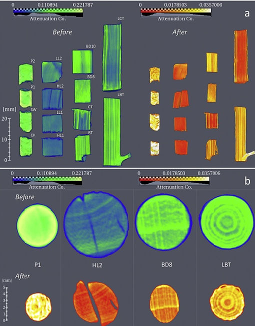
Graphical abstract from the paper "Torsion of a rectangular bar: Complex phase distribution in 304L steel revealed by neutron tomography", published in the journal Materials & Design in October 2022 by Khanh Van Tran, Robin Woracek, Nikolay Kardjilov, Henning Markötter, Daniel Abou-Ras, Stephen Puplampu, Christiane Förster, Dayakar Penumadu, Carl F.O.Dahlberg, John Banhart, Ingo Manke
https://doi.org/10.1016/j.matdes.2022.111037
This paper was published under the Creative Commons Attribution international Public License and permission for displaying this figure was given by Khanh Van Tran.
March 2023

Graphical abstract from the paper "Water flow through bone: Neutron tomography reveals differences in water permeability between osteocytic and anosteocytic bone material", published in the journal Materials & Design Volume 224 in December 2022 by Andreia Silveira, Nikolay Kardjilov, Henning Markötter, Elena Longo, Imke Greving, Peter Lasch, Ron Shahar, Paul Zaslansky.
https://doi.org/10.1016/j.matdes.2022.111275
This paper was published under the Creative Commons Attribution international Public License and permission for displaying this figure was given by Paul Zaslansky.
April 2023

FIGURE 3. Image analysis of the rat tibiae at different hydration states: dry—red, rehydrated 12 h—orange, rehydrated > 40 h—blue. (A) CNR for cortex vs. background, D2O vs. background, and D2O vs. cortex at the different hydration states, with zoom-ins of the first two hydration state for easier identification of the individual data points. Free D2O was only noticed in the third set of images of the two tibiae that were rehydrated for > 40 h (shown in B and C). (B) Transverse and (C) sagittal slices of tibia with regions for line profiles indicated with dashed square. Cortex, soft tissue, and D2O are indicated with white arrows. (D) and (E) Zoom-in of the regions in B and C, respectively, with corresponding separated and overlaid line profiles. Edges used for edge sharpness analysis are indicated with colored squares (cortex—purple, soft tissue—red). In the overlaid line profiles, thickening of the cortex with increased hydration is indicated with dashed lines. The arrows indicate increased signal due to D2O (solid), and reduced contrast for inner structures (dashed). The yellow dashed arrow indicates reduced contrast for inner structures after 12 h of rehydration.
Figure from the paper "The Hydration State of Bone Tissue Affects Contrast in Neutron Tomographic Images" by Elin Törnquist, Sophie Le Cann, Alessandro Tengattini, Lukas Helfen, Joeri Kok, Stephen A. Hall and Hanna Isaksson, published in Frontiers of Bioengineering and Biotechnology 2022; 10: 911866. doi: 10.3389/fbioe.2022.911866
This paper was published under the Creative Commons Attribution international Public Licence and permission for displaying this figure was given by Elin Törnquist.
May 2023

Figure 2. Neutron scattering images of charged and discharged, fresh and worn 90 mAh batteries. In the top row images, ξ = 2.25 μm. In the middle row images, ξ = 2.81 μm. At the bottom, plots show scattering values across the batteries. The color bar represents the dark-field signal acquired through each battery. The reported uncertainty is the one-sigma root mean square uncertainty of the nonlinear least squares fit.
Figure from the paper "Intact, Commercial Lithium-Polymer Batteries: Spatially Resolved Grating-Based Interferometry Imaging, Bragg Edge Imaging, and Neutron Diffraction" by Adam J. Brooks, Daniel S. Hussey, Kyungmin Ham, David L. Jacobson, Ingo Manke, Nikolay Kardjilov, Leslie G. Butler published in Appl. Sci. 2022, 12(3), 1281. https://doi.org/10.3390/app12031281
This paper was published under the Creative Commons Attribution international Public Licence and permission for displaying this figure was given by Les Butler.
June 2023

Figure 5. A 3D map of the patina thickness of the vase is shown in the planes xy (a), zy (b), and yx (c). The volume of the patina is colour-coded according to the thickness of the material, as expressed by the scale on the top-left corner. The vase is rendered with a semi-transparent surface. (d) The distribution of the thickness of the patina in the histogram shows a bimodal distribution.
Figure from the paper "An Insight into a Shang Dynasty Bronze Vessel by Nuclear Techniques" by Filomena Salvemini, Zeljko Pastuovic, Attila Stopic, Min-Jung Kim, Sue Gatenby published in Appl. Sci. 2023, 13(3), 1549;
https://doi.org/10.3390/app13031549
This paper was published under the Creative Commons Attribution international Public Licence and permission for displaying this figure was given by Filomena Salvemini.
